Complex and modified mouthparts in Histiostomatidae mites
Mites represent arachnids, which means that they share characters with much bigger organisms, such as spiders, skorpions or harvestmen. Their bodies consist of specialized bundles of segments, named tagmata. Two major tagmata are differed from each other in arachnids: prosoma, including legs and mouthparts, and opisthosoma, including for example the digestive and the reproductive systems.
Discussed diphyletic origin of mites
Mites are according to some acarological scientists eventually not longer just mites. The former two clades of mites, Parasitiformes and Acariformes, originally considered as sister taxa, were in some modern systematics reconstructed to be diphyletic. That would mean, there was no commor ancestor, from which only those two clades derived, the two major clades would be polyphyletic with no close relationship between them, each clade is assumed being closely related to different groups of arachnids (e.g. Psedoscorpions and Opiliones). Thus, when I talk about mites, I am talking about the clade Acariformes.
Mites of the Acariformes and body plan
In these Acariformes mites, the arachnid body construction plan was modified into three visible tagmata: gnathosoma (bearing chelicerae and pedipalps as mouthparts), proterosoma (bearing first two leg pairs) and hysterosoma (bearing last two leg pairs and opisthosoma organs).
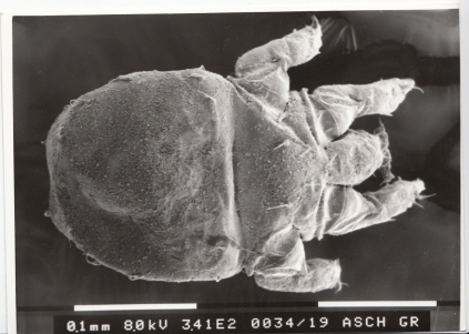
Male (large morph) of mite Histiostoma feroniarum in dorsal view. Body division in gnathosoma, proterosoma and hysterostoma. Fixation : critical-point-dried, SEM photography, copyrights Stefan F. Wirth
Mouthparts
Let’s talk about mouthparts, as they are an important aspect of my systematic and my function.morphological studies. Originally the gnathosoma consists of a pair of scissor-shaped chelicerae to grasp the food particles and of a pair of leg-shaped pedipalps, which mostly have mechano-sensitive and chemo-sensitive functions. But because mites colonized almost all kinds of existing habitats on earth, they extensively were exposed to the mechanisms of evolution. Acariform mites show a high range of variability regarding their morphology and their life strategies.
Mouthparts of Sarcoptiformes
Within the clade Sarcoptiformes, consisting of oribatid mites, Endeostigmata (seemingly paraphyletic) and astigmatid mites, there evolved a tendency towards miniaturization. Mites of the Astigmata are usually much smaller than one mm. Correspondingly the cuticle became thinner and softer, perfect adaptations to a life inside very tiny micro habitats, but at the same time also a limitation, namely towards more or less moist habitats due to the lack of a well developed desiccation protection. They appear inside compost, rotting wood or mammal dung, being even there very specifically adapted into very defined micro climatic conditions. They live in a world of complete darkness, which is why light sensory organs are completely lost or reduced to vestigial structures.
Inside their habitats, astigmatid mites need to reproduce, to develop through different nymphal stages until adulthood and of course to feed. Astigmata are no fluid suckers, but feed on particles, such as bacteria, algae, fungi, thus many Astigmata taxa can be named microorganism feeders.
Life-strategy of mites of the (family) Histiostomatidae

Extinct bark beetle fpssil in amber (collection Hoffeins) with phoretic mite deutonymphs. Fixation with hexamethyldisilazane, stereomicroscopic photography, copyrights Stefan F. Wirth
One of the largest family within the Astigmata clade is the Histiostomatidae, which I use since many years as model for my scientific studies. These mites are scientifically interesting from different points of view. Their ecology is characterized by life styles, which correspond to the life cycle of insects and other arthropods, to which most species have a close association. Most important aspect of these interactions between mites and other arthropods, commonly insects, is a dispersal strategy named „phoresy“. Mites use their „partners“ as carriers from one habitat to another. These habitats can often be the nests of the corresponding arthropods/ insects.
Habitats, in which mites of the Histiostomatidae develop successfully need to be moist and need to contain a sufficiant amount of microorganisms as food source. It is the most conspicuous feature of these mites to possess remarkably modified mouthparts compared to the above described standard equipment of an acariform gnathosoma.
Mouthparts of the Histiostomatidae
Mite Histiostoma sp. (sapropel around ponds, female, Berlin) feeding from a substrate surface inside its original habitat. Videography in 4K, copyrights Stefan F. Wirth
The character conditions of the gnathosoma were one of the reasons, why I at the beginning of my phd thesis in 2000 decided to put my research focus on this mite family, being worldwide in major still unexplored.
The chelicera modified into a dagger-like structure being formed by the fixed part of the former scissor-like organ, named the digitus fixus. There is a variability of shapes of this digitus fius-chelicera-ending within the Histiostomatidae . It can appear „simple-dagger-like, simple formed with a hook-like ending or having cuticular dentations of specific numbers and sizes along the lower edge of the digitus fixus.
As typical for mites of the big clade Astigmata, the pedipalps are reduced in size and almost immovably ventrally and dorsally connected with each other. In Histiostomatidae, the third pedipalp article is additionally distinctly bent sidewards. Their front sides bear more or less complex arrangements of flexible membraneous structures, which can morphologically differ between taxa or even species, thus giving them a systematic relevance. I named these membrane-organs „palparmembrane“ following the nomenclature, introduced by R. Scheucher in 1957. These membranes can be devided into fringes or being lobe-sphaped and can cover the last pedipalp article dorsally and/or ventrally. My histological analysis from 2006 indicated that these membranes are shaped by the enditesof the pedipalpal coxae.
Complex mouthpart apparatus
Thus Histiostomatidae possess a bizarre mouthpart apparatus being unique within the Acariformes and representing an amount of characters, which from the phylogenetc point of view can be reconstructed to have evolved in the stem species of that family (so called apomorphies).
Mouthpart apparatus as multifunctional organ


Mite Histiostoma sp. (male left, female right) feeding from a substrate surface inside its original habitat. Fixation with hexamethyldisilazane, SEM photography, copyrights Stefan F. Wirth
This gnathosoma is a multifunctional organ with the main function to select specific microorganism particles out of their liquid environments. When observing a histiostomatid mite with a sufficient high magnification walking along on a smooth water agar surface, on which bacteria and fungi growth was stimulated before, then occasionally trails can be seen around the walking mite, indicating that the gnathosoma was hold mostly leaned downwards towards the ground, pushing the microorganism cover along in front of the mite’s body. I interpreted this as an accumulation of food in order to gain more nutrients all at once. In my early papers, I described this as the typical feeding behavior of histiostomatid mites with the membraneous appendages acting like rubber sliders in the meantime. But as newer analyses showed is that such observations do not describe the full equipment of possible applications of the mite’s complex filter-feeding apparatus.
Membraneous structures create an underpressure to incorporate food


Mite Histiostoma ruehmi mouthpart endings with palparmembrane in ventral view. Fixation with hexamethyldisilazane, SEM photography, copyrights Stefan F. Wirth
More recent experiments with a higher videographic resolution and more suitable light conditions than 10 years ago (through-light and up light or one of them depending on the setting) showed that the palpar membrane structures , which more or less surround the entire fore-part (anterior part) of the gnathosoma can act like suckers: When the mite presses its front end of the mouthparts to the underground, an underpressure can be formed based on these membraneous structures. This seemingly facilitates the incorporation of nutrients in that area.
Note from January 2020: In retrospect, I do not consider it sensible to superficially describe the feeding behavior using the palpar membrane at the edge. A precise videographic analysis of individual images exists and is currently being developed into a scientific paper.

Aspects of the histiostomatid feeding behavior, including using the membranous components at the anterior end of the mouthparts (pedipalps), can partly be seen in the video below.
https://www.youtube.com/watch?v=fHliP_eHSGg
Mite Histiostoma ruehmi and an undetermined species feeding from a smooth artificial substrate surface and performing an underpressure to incorporate food. Videography, copyrights Stefan F. Wirth
Scanning-electron-microscopic experiments
 Mite Histiostoma cf feroniarum feeding in its original substrate, fixed with hexamethydisilazane, SEM
  copyrights Stefan F. Wirth


Mite Bonomoia opuntiae feeding from the surface of a substrate mount inside its original habitat. Rounded particles might represent yeast bodies. Fixation with hexamethyldisilazane, SEM photography, copyrights Stefan F. Wirth
In my early postdoc-years, still at the FU Berlin, I performed experiments in order to fix mite activities inside their original substrates by filling such a mite-substrate-setting up with 1,1,1,3,3,3-hexamethyldisilazane and warming the corresponding small experimental dish, until the chemical was vaporized. I then sputtered the conserved setting with gold and studied the details on it via scanning-electron-microscopy. Occasionally, mites were shrinkled or deformed after this procedure, but sometimes they stayed in shape and did seemingly still remain in their last activity positions. I several times could take SEM photos, showing that (well visible only in adult mites due to their size) mite specimens can insert their (distal) chelicerae-endings into bigger heaps of substrate (obviously full of nutrients) and use the entire laterally bent pedipalpal articles, including the connected palparmembranes, to lean it against the substrate surface, either to stabilize the chelicerae movents or even to support the incorporation of nutrients again by forming a slight underpressure, or both.
Mite species Bonomoia opuntiae
Early observations during times of my phd-thesis on the mite Bonomoia opuntiae could show that the mouthpart apparatus of this terrestrial/semiaquatic mite works well also under water or inside a watery juce of decomposing cactus pieces. There even a filter function comparable with a fishing net was hypothesised, but so far was never studied in detail. The very distinct fringes along the palparmembrane lobes in this mite species might support this theory. I also studied the semiaquatic mite Sarraceniopus nipponensis feeding inside watery environments (normally the digestive fluids of Sarracenia pitchers), again never focussing in detail in how excactly the feeding mechanism works.
A putatively new species
The herewith presented video shows behaviors of a female of the putative new species Histiostoma sp. , which I discovered in beginning of 2019 in sapropel around ponds inside an old gravel pit area in the Berlin forest Grunewald. The footage is presented in slow motion. The question was about how motile the whole gnathosoma apparatus in a histiostomatid species can be and what kinds of movements occured. As the settings, which I in early years of my mite studies used for videographic studies, were simplyfied and thus unnatural (smooth agar surfaces), I thought it being necessary and important to capture behaviors in a complexly sculptured habitat, namely surfaces of decomposing potato pieces (on which most histiostomatid species use to develop well).
It was visible, based on the specimens of my video of this species, that histiostomatid mites can be able to lift up their entire gnathosomas on a sometimes even higher position than the levels of the rest of their bodies. Additionally the gnathosoma can be turned to the right and to the left. Up and down as well as sideward movements of the whole feeding apparatus were often performed and represented obviously flexible reactions of the mite to the surface structure of the substrate and to the availability of suitable nutrients. In this context I was also interested in details of the movements of the chelicera tips themselves.
Chelicera endings (digitus fixus)
Although they can be used dagger-like and be accurately inserted into muddy substrate mounts, chelicera tips will also appear in a very fragile and seemingly careful way, when palpating the surface of the substrate underneath. Such chelicera movements are visible in the footage of this video, presented in slow motion (about 25 percent of original speed) and in a digital magnification. I interpret this visible fragility caution of the chelicerae as one option to discover suitable food sources. Other important organs perceive the mite’s environment chemically, modified setae, namely the so called solenidia, which might additionally recognize profitable microorganism sources.

Mite Histiostoma sp. (H. feroniarum-complex) feeding from substrate mounts inside its original habitat (A-F). Rounded particles might represent yeast bodies. D = distal chelicera endings (digitus fixus), holding food particles, fixation with hexamethyldisilazane, SEM photography, copyrights Stefan F. Wirth
Berlin, September 2019
Copyrights Stefan F. Wirth
